


Structure and Function of the Nucleolus
See Alberts et al, Molecular Biology of the Cell, "The Nucleolus is a Ribosome-Producing Machine", Garland Pub., 1994, pp 379-382.
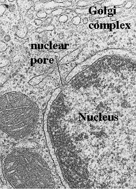
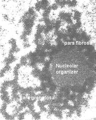
The nucleolus is organized from the "nucleolar organizing regions" on different chromosomes. A number of chromosomes get together and transcribe ribosomal RNA at this site. The nuclear organizing (NO) regions are seen as circular areas (pale) surrounded by a rim of electron dense filaments. These filaments collectively are called the pars fibrosa (PF). This is formed from newly transcribed ribosomal RNA. This diagram shows the parts of the nucleolus.
After the ribosomal RNA is transcribed, it is linked to proteins and one can see accumulation of ribonucleoprotein particles in the pars granulosa (PG). These particles form the two types of ribosomal subunits (large and small) which are then transported out of the Nuclear Pores separately. The pores do not accommodate the assembled ribosomes, therefore, they cannot reenter. This means that translation of RNA and synthesis of proteins must occur outside the nucleolus. For more information follow the link to ribosomes
After the ribosomal subunits are in the cytoplasm, they are connected and then, they may attach to endoplasmic reticulum . They provide an attachment and "code reading" site for the messenger RNA. If they attach to rough endoplasmic reticulum, they form a pore that moves the newly synthesized proteins into the cisterna of the rough endoplasmic reticulum .
The above figure shows another electron micrograph of a nucleolus with the nuclear organizing region, the pars fibrosa and the pars granulosa. Nucleoli increase in number and enlarge when the cell is stimulated to produce proteins. This is a sign that the cell has been stimulated or is actively involved in protein synthesis. Nucleoli disappear during cell division and then reform at the chromosomal nucleolar organizing centers.
Taken from Bloom and Fawcett, A Textbook of Histology, Chapman and Hall, 1994, Figure 1-15

One can study synthesis of ribosomal RNA with the electron microscope. A preparation of nucleoli is dissociated and spread on a liquid surface (in a manner similar to the spreading of chromatin). Then, the synthesis of ribosomal RNA is stimulated and after a period of time, the DNA from the nucleolar organizing region begins to look like a Christmas tree. The top of the tree is the start site. As you move down the "tree", the branches appear longer. Each branch is a growing strand of ribosomal RNA. The DNA code is being transcribed and the nucleotides added to the growing RNA strand.
Taken from Bloom and Fawcett, A Textbook of Histology, Chapman and Hall, 1994, Figure 1-17. For more information about the process, see the web site describing ribosomal functions.
For more information, contact:
Gwen Childs, Ph.D.,FAAA
Professor and Chair
Department of Neurobiology and Developmental Sciences
University of Arkansas for Medical Sciences
Little Rock, AR 72205
For questions, contact this email address:
